Medical researchers use laboratory-grown human cells to learn the intricacies of how cells work and test theories about the causes and treatment of diseases. The cell lines they need are “immortal”—they can grow indefinitely, be frozen for decades, divided into different batches and shared among scientists. In 1951, a scientist at Johns Hopkins Hospital in Baltimore, Maryland, created the first immortal human cell line with a tissue sample taken from a young black woman with cervical cancer. Those cells, called HeLa cells, quickly became invaluable to medical research—though their donor remained a mystery for decades. In her new book, The Immortal Life of Henrietta Lacks, journalist Rebecca Skloot tracks down the story of the source of the amazing HeLa cells, Henrietta Lacks, and documents the cell line's impact on both modern medicine and the Lacks family.
Who was Henrietta Lacks?
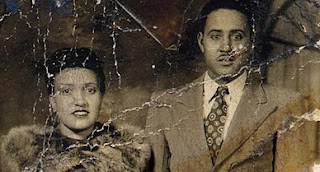
She was a black tobacco farmer from southern Virginia who got cervical cancer when she was 30. A doctor at Johns Hopkins took a piece of her tumor without telling her and sent it down the hall to scientists there who had been trying to grow tissues in culture for decades without success. No one knows why, but her cells never died.
Why are her cells so important?
Henrietta’s cells were the first immortal human cells ever grown in culture. They were essential to developing the polio vaccine. They went up in the first space missions to see what would happen to cells in zero gravity. Many scientific landmarks since then have used her cells, including cloning, gene mapping and in vitro fertilization.
There has been a lot of confusion over the years about the source of HeLa cells. Why?
When the cells were taken, they were given the code name HeLa, for the first two letters in Henrietta and Lacks. Today, anonymizing samples is a very important part of doing research on cells. But that wasn’t something doctors worried about much in the 1950s, so they weren’t terribly careful about her identity. When some members of the press got close to finding Henrietta’s family, the researcher who’d grown the cells made up a pseudonym—Helen Lane—to throw the media off track. Other pseudonyms, like Helen Larsen, eventually showed up, too. Her real name didn’t really leak out into the world until the 1970s.
How did you first get interested in this story?
I first learned about Henrietta in 1988. I was 16 and a student in a community college biology class. Everybody learns about these cells in basic biology, but what was unique about my situation was that my teacher actually knew Henrietta’s real name and that she was black. But that’s all he knew. The moment I heard about her, I became obsessed: Did she have any kids? What do they think about part of their mother being alive all these years after she died? Years later, when I started being interested in writing, one of the first stories I imagined myself writing was hers. But it wasn’t until I went to grad school that I thought about trying to track down her family.
How did you win the trust of Henrietta’s family?
Part of it was that I just wouldn’t go away and was determined to tell the story. It took almost a year even to convince Henrietta’s daughter, Deborah, to talk to me. I knew she was desperate to learn about her mother. So when I started doing my own research, I’d tell her everything I found. I went down to Clover, Virginia, where Henrietta was raised, and tracked down her cousins, then called Deborah and left these stories about Henrietta on her voice mail. Because part of what I was trying to convey to her was I wasn’t hiding anything, that we could learn about her mother together. After a year, finally she said, fine, let’s do this thing.
When did her family find out about Henrietta’s cells?
Twenty-five years after Henrietta died, a scientist discovered that many cell cultures thought to be from other tissue types, including breast and prostate cells, were in fact HeLa cells. It turned out that HeLa cells could float on dust particles in the air and travel on unwashed hands and contaminate other cultures. It became an enormous controversy. In the midst of that, one group of scientists tracked down Henrietta’s relatives to take some samples with hopes that they could use the family’s DNA to make a map of Henrietta’s genes so they could tell which cell cultures were HeLa and which weren’t, to begin straightening out the contamination problem.
So a postdoc called Henrietta’s husband one day. But he had a third-grade education and didn’t even know what a cell was. The way he understood the phone call was: “We’ve got your wife. She’s alive in a laboratory. We’ve been doing research on her for the last 25 years. And now we have to test your kids to see if they have cancer.” Which wasn’t what the researcher said at all. The scientists didn’t know that the family didn’t understand. From that point on, though, the family got sucked into this world of research they didn’t understand, and the cells, in a sense, took over their lives.